Department Introduction
Founded in November 1980, the Department of Radiology completed the construction of the picture archiving and communication system (PACS) in 2002, opening a new page for imaging diagnosis.
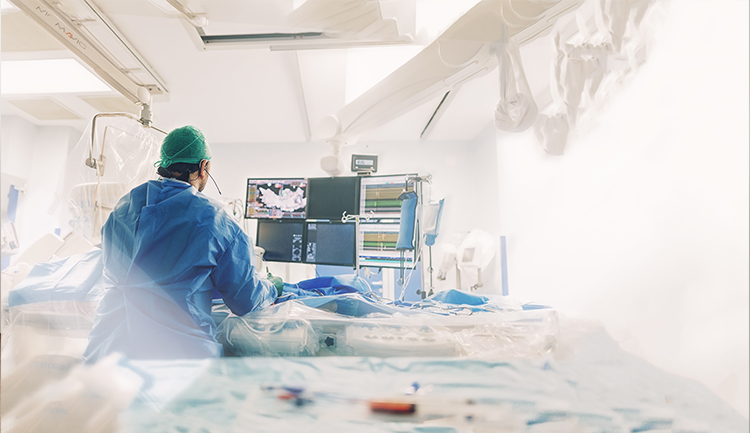
According to instrument characteristics and human organ systems, the Department of Radiology is divided into the General Radiology Division (chest radiation, skeletal joint radiation, pediatric radiation, abdominal imaging), Neuroradiology Division, Interventional Radiography Division, and so on.

The Department of Radiology currently owns a total of 47 instruments, including seven MRI scanners (five 1.5T and two 3T), six CT scanners (one 640-slice and five 128-slice), five digital X-ray fluoroscopes, 12 digital medical X-ray radiography systems, seven mobile X-ray systems, two absorptiometry machines, two DR mammography machines, and six ultrasound scanners.
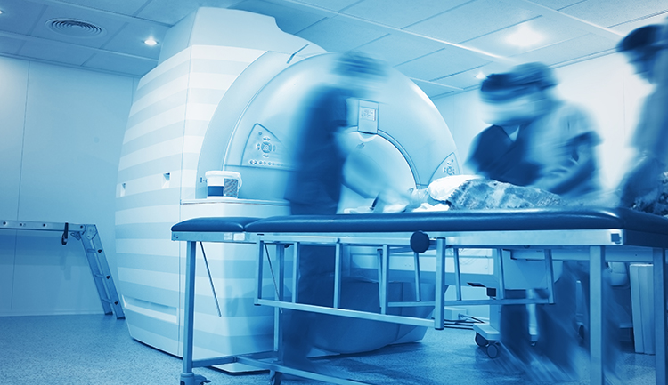
Magnetic Resonance Imaging (MRI) uses radio frequency (RF) waves to stimulate hydrogen atoms in the body's water and fat to produce resonance, thus giving rise to an image generated by a signal of different intensity.
Computed Tomography (CT) uses a large number of X source rays to penetrate the human body to obtain images. A two-dimensional (2D) image made by a computer is used to observe the interior of the body. In recent years, with the rapid development of science and technology and computers, CT has entered multi-layer tomography. Combined with the powerful image function of the computer workstation that can produce three-dimensional (3D) images, more CT angiography and virtual colonoscopies and tracheoscopies can be made. Almost all parts of the body can be checked with CT.
Bone density scanning uses dual-energy X-ray to scan the examined site, and then the scintillation detector receives the X-ray that penetrates the detected site. The data obtained from computer analysis are used to calculate bone density, which is generally being applied to the detection of osteoporosis.
Ultrasonography utilizes a high-frequency sound wave that cannot be heard by the human ear, which travels through the body to the inside and the refracted sound wave is transformed by the instrument to display the image for examining the shape, size, position, and movement of the organs and tissues of the human body. Currently, it is often being applied to the scanning of the pelvic cavity, the heart vessels, the abdomen, and some superficial tissue organs.


 Switch
 Homepage
 facebook
 IG
IG
 Other Hospital
 CMUH App


 Facebook
 IG
IG
 YouTube